


Lumbar osteochondrosis is a chronic disease that develops as a result of a degenerative-dystrophic process on the intervertebral discs. The disease is widespread and affects most people between the ages of 25 and 40.
According to statistics, every second adult experiences back pain at least once in their life, while in 95% of cases it is caused by osteochondrosis of the spine.
Patients with a severe course of lumbar osteochondrosis, with persistent pain and other manifestations are recognized as temporarily incapacitated. If their condition does not improve within four months, the issue of establishing a disability group is decided.
Lumbar osteochondrosis is a serious medical and social problem, because the disease mainly affects people of the youngest age, and in addition, in the absence of treatment, it can cause the formation of a herniated disc.
Causes and risk factors
Factors that predispose to the development of lumbar osteochondrosis are:
- spinal structure anomalies;
- lumbarization - congenital pathology of the spine, characterized by the separation of the first vertebra from the sacrum and its transformation into the sixth (additional) lumbar;
- sacralization is a congenital pathology in which the fifth lumbar spine is fused with the cross;
- asymmetric arrangement of the intervertebral joints;
- pathological narrowing of the spinal canal;
- reflects spondiogenic pain (somatic and muscular);
- obesity;
- sedentary lifestyle;
- prolonged exposure to vibration;
- systematic physical stress;
- smoking
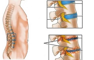
Unfavorable static-dynamic loads in combination with one or several risk factors lead to a change in the physiological properties of the pulpal disc of the fibrous disc, which plays the role of shock absorption and ensures the mobility of the spine. This process is based on the depolymerization of polysaccharides, which leads to the loss of moisture in the tissue of the gelatinous nucleus. As a result, the nucleus pulposus, and with it the fibrous disc, loses its elastic properties. Further mechanical stress causes the bulging of the annular fibrosus to lose elasticity. This phenomenon is called bulging. Cracks appear on the fibrous nucleus through which fragments of the pulposus nucleus (prolapse, disc herniation) fall out.
Prolonged compression of the nerve roots that innervate certain organs of the abdominal cavity over time leads to a deterioration in their functioning.
Instability of the spinal segment is accompanied by reactive changes in the bodies of adjacent vertebrae, intervertebral joints and accompanying spondyloarthritis develops. Significant muscle contraction, for example, in the background of physical activity, leads to displacement of the vertebral body and entrapment of nerve roots with the development of radicular syndrome.
Another cause of pain and neurological symptoms in lumbar osteochondrosis can be osteophytes - the growth of bones on the processes and bodies of the vertebrae that cause radicular syndrome or compression myelopathy (compression of the spinal cord).Forms of the disease
Depending on which structures are involved in the pathological process, lumbar osteochondrosis is clinically manifested by the following syndromes:
- reflex- lumbodinia, lumboischalgia, lumbago; develop against the background of reflex overexertion of the back muscles;
- compression (spinal, vascular, radicular)- compression (compression) of the spinal cord, blood vessels or nerve roots leads to their development. Examples are lumbosacral radiculitis, radiculoischemia.
Symptoms of lumbar osteochondrosis
In lumbar osteochondrosis, the symptoms are determined by which structures are involved in the pathological process.
Lumbago occurs under the influence of hypothermia or physical overexertion, and sometimes for no apparent reason. The pain appears suddenly and is of a shooting nature. It is intensified by sneezing, coughing, turning the body, exercising, sitting, standing, walking. In the supine position, the sensations of pain are significantly weakened. Sensitivity and reflexes are preserved, the range of motion in the lumbar spine is reduced.
Observe by palpation:
- pain in the lumbar region;
- paravertebral muscle spasm;
- smoothing of lumbar lordosis, which in many cases is combined with scoliosis.
The nerve root tension syndrome in the lumbago is negative. When they lift a straight leg, patients notice an increase in pain in the lumbar region, rather than their appearance in the elongated lower limb.
Pain attacks often occur in lumbar osteochondrosis, becoming more intense and long-lasting each time.
In lumbodinia, the clinical picture resembles lumbago, but the increase in pain intensity occurs over several days.
With lumboischalgia, patients complain of pain in the lumbar region radiating to one or both lower extremities. The pain spreads to the buttocks and back of the thighs and never reaches the feet.
Lumboischalgia is characterized by vasomotor disorders:
- changes in temperature and skin color of the lower extremities;
- feeling hot or cold;
- violation of blood circulation.
The development of lumbar compression syndrome is clinically manifested by the following symptoms:
- dermatomal hypalgesia;
- shooting pains;
- weakening or complete loss of deep reflexes;
- peripheral paresis.
In compression syndromes, the pain is aggravated by bending the torso, sneezing and coughing.
Diagnostics
The diagnosis of lumbar osteochondrosis is made on the basis of clinical picture of the disease, laboratory and instrumental research methods.
In blood tests for lumbar osteochondrosis:
- decrease in calcium concentration;
- increased ESR;
- increased alkaline phosphatase levels.
In the diagnosis of lumbar osteochondrosis, great importance is attached to the X-ray examination of the spine.
Prolonged compression of the nerve roots that innervate certain organs of the abdominal cavity over time leads to a deterioration in their functioning.
X-ray signs confirming the diagnosis are:
- change the configuration of the affected segment;
- pseudospondylolisthesis (movement of adjacent vertebral bodies);
- deformation of the closing plates;
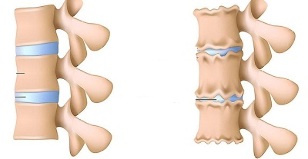
- alignment of the intervertebral disc;
- uneven height of the intervertebral disc (symptom of spacing), which is associated with asymmetric muscle tone.
Also used in the diagnosis of lumbar osteochondrosis, if indicated, are:
- myelography, computed tomography or magnetic resonance imaging - are necessary for permanent symptoms, the development of neurological deficits;
- scintigraphy (study of phosphorus accumulation in the skeletal system, marked with tech-99) - is performed if there is a suspicion of a tumor or infectious process, spinal cord injury.
The differential diagnosis of lumbar osteochondrosis is made with the following diseases:
- spondylolisthesis;
- dyshormonal spondylopathy;
- ankylosing spondylitis (ankylosing spondylitis);
- infectious processes (disc inflammation, osteomyelitis of the spine);
- neoplastic processes (primary tumor of the spine or its metastatic lesions); rheumatoid arthritis;
- deforming osteoarthritis of the hip joint;
- reflected pain (diseases of internal organs and large blood vessels).
Treatment of lumbar osteochondrosis
For lumbar osteochondrosis, the following treatment tactics are usually followed:
- bed rest 2-3 days;
- traction of the affected segment of the spine;
- strengthening the muscles of the back and abdomen (creating the so-called muscle corset);
- influence on pathological myofascial and myotonic processes.
Lumbago occurs under the influence of hypothermia or physical overexertion, and sometimes for no apparent reason.
In most cases, conservative treatment of lumbar osteochondrosis is performed, including the following measures:
- infiltrative anesthesia of muscles with a solution of local anesthetics;
- taking nonsteroidal anti-inflammatory drugs;
- taking desensitizing agents;
- vitamin therapy;
- taking tranquilizers and antidepressants;
- manual therapy, massage;
- physiotherapy exercises;
- acupuncture;
- postisometric relaxation.
The absolute indications for surgical treatment of lumbar osteochondrosis are:
- acute or subacute spinal cord compression;
- development of cauda equina syndrome, characterized by pelvic organ dysfunction, sensory and movement disorders.
Therapeutic exercises for lumbar osteochondrosis
Physical therapy plays a significant role in the complex treatment of lumbar osteochondrosis. Regular exercises allow you to normalize the muscle tone of the paravertebral muscles, improve metabolic processes in the tissues affected by the pathological process, and also form a well-developed muscular corset that can support the spine in the correct position, relieve unnecessary static loads.
In order for gymnastics with lumbar osteochondrosis to have the greatest effect, you need to adhere to the following principles:
- regularity of teaching;
- gradual increase in the intensity of physical activity;
- avoiding overwork during classes.
Physiotherapy should be performed under the guidance of an experienced instructor, who will select the exercises that are most effective for a particular patient and control the correctness of their implementation.
According to statistics, every second adult experiences back pain at least once in their life, while in 95% of cases it is caused by osteochondrosis of the spine.
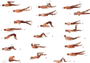
In addition to teaching with an instructor, you should perform a set of morning exercises every day, which includes special exercises for lumbar osteochondrosis.
- Relaxation and contraction of the abdominal muscles.Starting position is standing, feet shoulder-width apart, arms lowered along the body. Inhale smoothly, relaxing the muscles of the anterior abdominal wall. During exhalation, retract the abdomen as much as possible, tensing the abdominal muscles. The exercise should be repeated until mild fatigue occurs.
- Head movements with spinal flexion.The starting position is kneeling, leaning on the floor with arms outstretched, back straight. Slowly raise your head and bend at the back. Hold this position for a few seconds, then return smoothly to the starting position. Repeat at least 10-12 times.
- "Pendulum".Starting position lying on your back, arms close to the body, legs bent at right angles at the knees and hips. Turn your legs to the right and left with pendulum-like waving movements, trying to reach the floor. In this case, the blades cannot be torn off the floor.
- Ship.Starting position lying on your stomach, arms outstretched forward. Tear your upper body and legs off the floor, bending at the back. Hold this position for 5-6 seconds and slowly return to the starting position. Run 10 times.
Potential consequences and complications
The main complications of lumbar osteochondrosis are:
- formation of intervertebral hernia;
- vegetative-vascular dystonia; spondylolysis, spondylolisthesis; osteophytosis; spondyloarthritis;
- spinal canal stenosis, which leads to compression of the spinal cord and can cause permanent disability and reduced quality of life.
Prolonged compression of the nerve roots innervating certain organs of the abdominal cavity over time leads to a deterioration in their functioning. As a result, patients have bowel dysfunctions (constipation, diarrhea, bloating) and pelvic organs (urinary disorders, erectile dysfunction, frigidity, infertility).
Forecast
Pain syndrome in lumbar osteochondrosis occurs in the form of remission and exacerbation. Lumbago lasts 10-15 days, after which the patient's condition improves, the pain subsides. A favorable outcome can be prevented by associated secondary diseases. Often in lumbar osteochondrosis, attacks of pain are repeated, which become more and more intense and long-lasting each time.
Physical therapy plays a significant role in the complex treatment of lumbar osteochondrosis.
Patients with a severe course of lumbar osteochondrosis, with persistent pain and other manifestations are recognized as temporarily disabled. If their condition does not improve within four months, the issue of establishing a disability group is decided.
Prevention
Prevention of the development of osteochondrosis of the spine consists of the following measures:
- smoking cessation;
- weight normalization;
- improvement of general physical condition, active lifestyle;
- avoiding provocative conditions (lifting weights, sudden movements, turns, bending).